 By Dr. L. Claudia Pop, MD
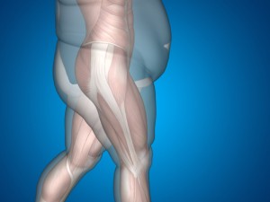
Osteoporosis and obesity represent increasing health burdens worldwide. Bone fragility and increased risk of fracture, resulting from bone loss and microscopic damage of the skeleton, constitute the hallmark of osteoporosis.1
BONE ACCRUAL
The major predictor for osteoporosis risk later in life is peak bone mass achieved during second decade of life. In adolescents and young adults, genes (~75% of an individual’s peak bone mass is genetically pre-determined), hormonal milieu and environmental factors interact to promote axial growth and skeletal expansion, mainly by stimulating bone formation.2 Conditions that alter bone formation and/or increase bone resorption and alterations in the hormonal cascade accompanying puberty (e.g. growth-hormone deficiency, delayed puberty) are associated with lower peak bone mass and increased risk of fracture later in life.1 Notably, higher adiposity in children prevents maximal bone mass accrual at certain sites of the skeleton and increases the risk of fracture in overweight/obese children.3
BONE LOSS
Starting from the 4th decade of life, there is a 0.3-0.5% yearly loss of bone mass in both genders due to higher rates of bone resorption compared to formation.4 In women, the dramatic drop in estrogen levels during early years of menopause is associated with a rapid decline in bone mass.5 In addition, certain medication could further enhance bone loss (e.g. glucocorticoids). In men, the decrease in sex hormones production starts later in life and is much slower compared to women, hence the decline in bone mass is delayed.6 In addition, independently of steroid decline, in both genders, aging causes lower bone formation and bone loss.7
OBESITY
On the other hand, obesity is defined as excessive adipose tissue accumulation and is associated with poor health status.8 A combination of causes and associated factors induce obesity. Among those: unhealthy dietary patterns, sedentary life style, medication and other exposures as well as genes are the most important. The amount of adipose tissue changes throughout life such that, with aging, percent body fat increases while lean mass and bone mineral density decrease.9 Moreover, as people age, a redistribution of the adipose tissue occurs, with excessive fat tissue being localized specifically in the abdominal area.10
Previously considered two distinct disorders, common features between osteoporosis and obesity have been noticed:
- Genetic and environmental factors are involved in the pathology of both diseases
- Both start early in life and their incidence increase with aging
- Bone formation cells (osteoblasts) and fat cells (adipocytes) share a common origin: the mesenchymal stem cell found within the bone marrow.
EVIDENCE
Although, obesity increases the risk for cardiovascular disease, hypertension and type 2 diabetes, excess body weight has been considered protective against osteoporosis and fractures due to higher bone mass in the obese.11-15Nevertheless, recent data show that obesity, particularly abdominal obesity, is inversely related to bone mineral density.16-18
Newly developed imaging techniques such as quantitative computed tomography (CT), which can measure the amount of adipose tissue present in the abdominal cavity (visceral fat) and the subcutaneous adipose tissue, suggest that intraabdominal fat depot is a strong predictor for lower BMD and compromised bone quality.16, 17, 19 In fact, epidemiological studies suggest that obese individuals may have a greater risk of fracture.20, 21
Furthermore, some clinical interventions have opposite effects on fat versus bone mass and support the deleterious effects of fat on bone and the inverse relationship between fat and bone mass. For example, physical activity decreases fat mass while increasing bone mass. Milk and tea consumption are thought to play a role in prevention of both osteoporosis and obesity.22
In addition, observational studies reported that high calcium intake may promote weight or fat loss, although this has not been confirmed by clinical trials.23 Accelerated bone loss, increased fat mass, and decreased lean mass accompany menopause and hormone replacing therapy effectively attenuates these changes.24 In some situation, osteoporosis and obesity could become major consequences of treatments. For instance, glucocorticoids have been shown to decrease bone mass and increase abdominal adiposity.25
MECHANISMS
Adipose tissue is not only a major energy storage depot; it has also been recognized as a major endocrine organ.26, 27A major hormone produced in the adipose tissue is leptin.28 Among many endocrine effects, leptin has been involved in bone metabolism. Studies conducted in leptin-deficient mice have shown increased bone mass despite higher cortisol and hypogonadism, both causing bone loss.
Moreover, increased death of osteoblasts upon exposure to leptin has been reported.29, 30 However, the effects of leptin on bone may differ with skeletal site. In addition to being a hormone reservoir, adipose tissue secretes several bioactive peptides, collectively named adipokines, which act both locally and systemically and affect bone metabolism.31
Also, adipose tissue releases inflammatory cytokines which can increase bone resorption and decrease bone mass.32, 33 On the other hand, the effect of subcutaneous fat on bone is somewhat controversial. Some reported beneficial effects of subcutaneous fat on bone possibly due to additional mechanical loading on bone or lower production of hormones and inflammatory markers compared to visceral fat while others have found deleterious effects.17, 34
Despite a certain level of uncertainty and controversy in the literature regarding the effects of body fat distribution on bone, the evidence that obesity may be related to an increased risk of fracture has been accumulating.
TYPES OF FAT
WHITE
In humans, excess energy is stored as white adipose tissue (WAT) which can be found dispersed throughout the body, either surrounding major organs or in subcutaneous localization such as buttocks or the thighs. In addition, WAT can be present in many other regions of the body as well as within the bone marrow. Depending on localization, adipose tissue may be receptive to steroid hormones such as sex hormones or glucocorticoids. Distribution of WAT tissue is largely controlled by genes.
BROWN
Aside from white fat – the major energy storage organ, mammals also possess brown adipose tissue (BAT) which is involved in energy expenditure in the form of thermogenesis or heat production. The presence of uncoupling protein-1 (known as UCP1), which dissipates the energy produced by cellular respiration into heat is a unique feature of brown adipose cell.35
Although, BAT is found primarily in fetuses and newborns and acts to regulate body temperature in response to cold, it becomes almost insignificant in adults and its roles are less clear. The decline of active BAT with aging has been proposed as a cause for a less efficient thermogenesis process and energy expenditure regulation in the elderly.36
In addition, a distinct group of brown fat cells, with thermogenic capacity can develop into white adipocytes in response to certain stimuli.37 These cells have been referred to as beige or “brite” (brown in white) adipocytes and seem to have a role in thermogenesis similar to brown fat cells. It is still unclear to what extent the adipose cells with thermogenic capacity identified in adult humans are brown or beige adipose cell.
THE ORIGIN OF ADIPOSE TISSUE
The bones forming the axial skeleton (skull, vertebrae and thoracic cage) contains bone marrow (or myeloid tissue), a highly vascular and a cell rich environment. Two types of cells are particularly important:
- mesenchymal stem cells that can generate bone cells (osteoblasts), fat cells (adipocytes-white/brown) and cartilage cells (chondrocytes)
- hematopoietic stem cells that can form blood cells.
In children, bone marrow is mainly red bone marrow containing hematopoietic cells and primarily found in long bones such as the femur. Throughout childhood, red bone marrow gradually becomes yellow, fatty bone marrow, while red bone marrow is limited to the skull, thorax bones, pelvis, and proximal femur. Beginning the forth decade of life, more than 50% of bone marrow cells are replaced by fat and almost all becomes fat in the elderly. Recent studies suggest that within the bone marrow environment, mesenchymal stem cells are also essential at maintaining hematopoietic stem cells.38
With the help of newly developed imaging techniques (Magnetic Resonance Imaging-MRI), cavities inside bones previously considered empty have been shown to contain fat even in healthy young people. Furthermore, in individuals with osteoporosis the amount of fat observed inside these cavities exceeds that found in healthy people. In addition, other factors associated with bone loss (i.e. diabetes, immunosuppressive medication, prolonged bed rest, ovariectomy) simultaneously cause fat accumulation in the bone.
Furthermore, visceral adiposity is associated with increased bone marrow fat and lower bone formation.39 The link between brown adipose tissue and bone is currently limited at animal models and is inconsistent, some studies showing a positive effect of brown adipocytes on bone remodeling while others showing increased brown adipogenesis at the expense of bone formation. However, as brown adipose tissue (BAT) is less apparent at older age at a time when thermoregulation is less efficient while bone loss is inevitable suggest a common background.
Characterizing the biochemical signals that modulate the ability of mesenchymal stem cells to differentiate into bone cells or adipose cells to possibly develop treatments targeting bone disease and other disorders has been an area of research developing rapidly in the recent years. Available drugs for osteoporosis are associated with adverse effects, therefore controlling mesenchymal stem cells differentiation into bone cells rather than fat could prevent or treat osteoporosis.
REFERENCES
- U.S. Department of Health and Human Services. The Surgeon General’s Report on Bone Health and Osteoporosis: What It Means To You. U.S. Department of Health and Human Services, Office of the Surgeon General, 2012.
- Recker RR and Heaney RP (1993) Peak bone mineral density in young women. JAMA270: 2926–2927.
- Goulding A et al. (2001) Bone mineral density and body composition in boys with distal forearm fractures: a dual-energy X-ray absorptiometry study. J Pediatr 139: 509–515.
- Manolagas SC. Birth and death of bone cells: basic regulatory mechanisms and implications for the pathogenesis and treatment of osteoporosis. Endocr Rev.2000;21:115–37.
- Lindsay R 1988 Sex steroids in the pathogenesis and prevention of osteoporosis. Osteoporosis: Etiology, Diagnosis and Management. Raven Press, New York, pp 333–358.
- Melton III LJ, Atkinson EJ, O’Connor MK, O’Fallon WM, Riggs BL. 1998 Bone density and fracture risk in men. J Bone Miner Res 13:1915–1923.
- Parfitt AM 1992 The two-stage concept of bone loss revisited. Triangle 31:99–110.
- http://www.who.int/topics/obesity/en/
- St-Onge MP, Gallagher D. Body composition changes with aging: the cause or the result of alterations in metabolic rate and macronutrient oxidation? Nutrition. 2010 Feb;26(2):152-5
- Atlantis E, Martin SA, Haren MT, Taylor AW, Wittert GA. Lifestyle factors associated with age-related differences in body composition: the Florey Adelaide Male Aging Study. Am J Clin Nutr. 2008;88:95–104.
- Nejat EJ , Polotsky AJ , Pal L. Predictors of chronic disease at midlife and beyond—the health risks of obesity. Maturitas. 65:106–111.
- De Laet C , Kanis JA , Oden A, et al.. Body mass index as a predictor of fracture risk: a meta-analysis. Osteoporos Int. 2005;16:1330–1338.
- Pittas A , Harris SS , Eliades M , Stark P , Dawson-Hughes B. Association between serum osteocalcin and markers of metabolic phenotype. J Clin Endocrinol Metab. 2009;94:827–832.
- Reid IR, Plank LD, Evans MC. Fat mass is an important determinant of whole body bone density in premenopausal women but not in men. J Clin Endocrinol Metab. 1992;75:779–782.
- Albala C , Yanez M , Devoto E et al. Obesity as a protective factor for postmenopausal osteoporosis. Int J Obes Relat Metab Disord. 1996;20:1027–1032.
- Bredella MA , Torriani M , Ghomi RH, et al.. Determinants of bone mineral density in obese premenopausal women. Bone. 2011;48:748–754.
- Gilsanz V , Chalfant J , Mo AO et al. Reciprocal relations of subcutaneous and visceral fat to bone structure and strength. J Clin Endocrinol Metab.2009;94:3387–3393.
- Pollock NK , Laing EM , Hamrick MW et al. Bone and fat relationships in postadolescent black females: a pQCT study. Osteoporos Int.2011;22:655–665.
- Choi HS , Kim KJ , Kim KM, et al.. Relationship between visceral adiposity and bone mineral density in Korean adults. Calcif Tissue Int. 2010;87:218–225.
- Nielson CM , Marshall LM , Adams AL, et al.. BMI and fracture risk in older men: the osteoporotic fractures in men study (MrOS). J Bone Miner Res. 2011;26:496–502.
- Compston JE , Watts NB , Chapurlat R, et al.. Obesity is not protective against fracture in postmenopausal women: GLOW. Am J Med. 2011;124:1043–1050.
- St Onge MP. Dietary fats, teas, dairy, and nuts: Potential functional foods for weight control. Am J Clin Nutr. 2005;81:7–15.
- Zemel MB. Role of calcium and dairy products in energy partitioning and weight management. Am J Clin Nutr. 2004;79:907S–912S.
- Manson JE, Martin KA. Postmenopausal hormone-replacement therapy. N Engl J Med. 2001;345:34–40.
- Zhao LJ, Jiang H, Papasian CJ et al. Correlation of obesity and osteoporosis: effect of fat mass on the determination of osteoporosis. J Bone Miner Res. 2008 Jan;23(1):17-29.
- Flier JS, Cook KS, Usher P, Spiegelman BM. Severely impaired adipsin expression in genetic and acquired obesity. Science 1987;237:405–408
- Siiteri PK 1987 Adipose tissue as a source of hormones. Am J Clin Nutr 45:277–282
- Zhang Y, Proenca R, Maffei M et al. Positional cloning of the mouse obese gene and its human homologue. Nature 1994;372:425–432
- Cock TA, Auwerx J 2003 Leptin: cutting the fat off the bone. Lancet 362:1572–1574
- Gordeladze JO, Drevon CA, Syversen U, Reseland JE. Leptin stimulates human osteoblastic cell proliferation, de novo collagen synthesis, and mineralization: Impact on differentiation markers, apoptosis, and osteoclastic signaling. J Cell Biochem. 2002;85:825–36
- Conde J, Scotece M , Abella V et al. Basic Aspects of Adipokines in Bone Metabolism. Clinic Rev Bone Miner Metab (2015) 13:11–19.
- Cao JJ. Effects of obesity on bone metabolism. J Orthop Surg Res. 2011;6:30
- Ibrahim SE, El Shishtawky HF, Helmy A, Galal ZA, Salam MHA. Serum leptin concentration, bone mineral density and bone biochemical markers in a sample of Egyptian women: a possible relationship.Egyptian Rheumatologist. 2011;33:171–177
- P. T. Katzmarzyk, T. V. Barreira, D. M. Harrington et al. Relationship between abdominal fat and bone mineral density in white and African American adults. Bone, vol. 50, no. 2, pp. 576–579, 2012
- Devlin MJ. The “Skinny” on brown fat, obesity, and bone. Am J Phys Anthropol. 2015 Feb;156 Suppl 59:98-115.
- Gesta S, Tseng YH, Kahn CR. Developmental origin of fat: tracking obesity to its source. Cell. 2007 Oct 19;131(2):242-56. Review. Erratum in: Cell. 2008 Oct17;135(2):366
- Vitali, A. et al. The adipose organ of obesity-prone C57BL/6J mice is composed of mixed white and brown adipocytes. J. Lipid Res. 53, 619–629 (2012).
- Frenette PS, Pinho S, Lucas D, Scheiermann C. Mesenchymal stem cell: keystone of the hematopoietic stem cell niche and a stepping-stone for regenerative medicine. Annu Rev Immunol. 2013;31:285-316.
- Bredella MA, Torriani M, Ghomi RH et al. Vertebral bone marrow fat is positively associated with visceral fat and inversely associated with IGF-1 in obese women. Obesity (Silver Spring). 2011 Jan;19(1):49-53.
I went over this web site and I conceive you have a lot of fantastic info, bookmarked (:.